By Oluebubechukwu Eze
Reactive oxygen species (ROS) are natural by-products of energy metabolism and are considered toxic molecules because of their high reactivity to most biological macromolecules, which generally include DNA damage, oxidation of polyunsaturated fatty acids (PUFA) and amino acids, and inactivation of some specific enzymes. Oxidative stress occurs in a cell or tissue when the concentration of ROSs exceeds the antioxidant capability of the cell. This page explores the negative effects ROS induced by sleep deprivation, as a result of skeletal muscle function, and as a cause of male infertility.Influence of ROS- Sleep deprivation and severe life stress
Oxidative stress is a common feature of numerous central nervous system (CNS) disorders. The accumulation of ROS in the CNS is known to increase the brain's susceptibility to tissue damage. They trigger numerous molecular cascades which lead to increased blood-brain barrier permeability (through activation of matrix metalloproteinases and subsequently degradation of tight junctions), alterations of brain morphology, neuroinflammation, and neuronal death (Gu et al., 2011).Sleep is a universal, dynamic brain process present in organisms ranging from invertebrates to mammals, which serves a homeostatic function. Our biological clock rhythm plays a crucial role in the development of several CNS functions, which include memory, learning, and neurogenesis. Prolonged sleep deprivation is a well known severe life stressor (SLS) and sometimes can result in irreversible oxidative damage to brain tissue. The necessary hours of sleep needed to rest brain activity varies among different organisms and within their respective age ranges. In an experiment performed by Cem et al., 2011, rodents with chronic sleep deprivation showed structural remodeling of brain regions (such as hippocampus, amygdala, and prefrontal cortex) which are involved in the formation and consolidation of memory. Brain antioxidant capacity was also strongly diminished in sleep-deprived animals (Carol et al., 2005, Ramanathan et al., 2002) with subsequent reduced glutathione levels and superoxide dismutase activity in the brain.
In another study performed by Carol et al., 2005, chronic administration of Vitamin E, a strong antioxidant, can reverse the effects of chronic sleep deprivation-induced cognitive impairment. Vitamin E is able to normalize activities of catalase, SOD, and glutathione peroxidase.
Impact of Oxidative Stress on Skeletal Muscle
Skeletal muscle is a highly specialized tissue in the body that functions with excellent plasticity when responding to external stimulus such as exercise and the 'fight or flight response.' Excessive muscle activity such as during exercise promotes ROS production in the mitochondria; and the activities of NADPH oxidase, Xanthine oxidase, myostatin, and phospolipase A2 produce varying levels of ROSs as well. Research by Jonathan et al., 2012 showed that ROS levels are increased in subjects with aging-related sarcopenia, cardiac reperfusion injuries or muscular diseases.
Effects of ROS on Force Generation and Muscular Atrophy
During normal physiological conditions of skeletal muscle, low-level intracellular ROS are essential for normal (proper function of motor proteins in muscle fibers) force generation. Although exercise generally increases muscle strength and mass, in individuals who are not physically fit and live sedentary lifestyles, a single bout of exhaustive exercise has been shown to cause oxidative damage to muscular tissue. The same does not hold, however, for individuals who are physically fit and actively train themselves due to their increased resistance to oxidative stress. Exercise demands a lot energy which is supplied by mitochondria in muscle cells, and an increase in skeletal muscle mitochondrial activity results in a corresponding increase of ROSs. Figure 1 shows some deleterious and beneficial effects of exercise-induced ROS increase. The effects are also dependent on the concentration of ROS and duration of ROS exposure in the individual. Strong increases in ROS after an intense workout, during aging or a disease state can cause contractile dysfunction and muscle atrophy, which results in muscle weakness and fatigue. It is important to note, however, that the amount of ROS produced during exercise varies within individuals.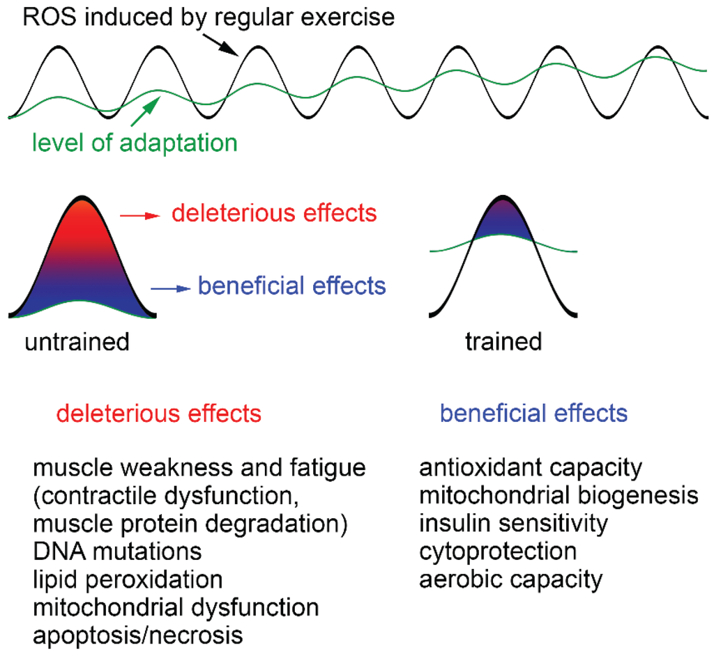
Contractile Dysfunction
Heart failure is charactrized by contractile muscle dysfunction and a high incidence of sudden death from nonreentrant ventricular arrhythmias, both of which involve altrered intracellular calcium handling (Pogwizd and Bers, 2002). Muscle contraction depends on sets of following sequences of events referred to as excitation-contraction coupling. Action potentials are stimulated at the neuromuscular junction and then propagated along the surface membrane of a muscle fiber and into the transverse tubular system where it is sensed by dihydropyridine receptors (DHPRs). DHPR activation triggers the opening of Ca2+ release channels (also known as ryanodine receptors) in the sarcoplasmic reticulum, which causes an increase in cytoplasmic free calcium (Hidalgo et al., 2006). There is some uncertainty surrounding the effects of ROS on the force generating myofilaments, however, it is generally accepted that long exposure to hydrogen peroxide results in force decline. Steinbacher and Eckl, 2015, suggested the variations in generated force were the result of changes in the myofibrillar Ca2+ sensitivity. Troponin I, which is involved in sensing intracellular Ca2+ levels is a target of ROS. Cysteine residues in the active site of the protein react with glutathione to protect it from oxidative stress and increase the Ca2+ sensitivity of the contractile apparatus (Mollica et al., 2012).
Muscle Atrophy
Reactive oxygen species are also capable of of modulating signalling pathways (Fig. 2 shows the effect of ROS in a diseased muscle or one without regular exercise) like calcium, protein tyrosine kinases and phosphatases, serine/threonine kinases, and phospholipases. This implicates ROS in their involvement with changes in gene expression, cell function, metabolism or cell damage. Jonathan et. al., 2012 in their study, discovered high ROS levels which caused sustained activation of NF- kB and FoxO pathways that then activated two muscle-specific E3 ubiquitin ligases, atrogin-1 or muscle atrophy F-box (MAFbx) and muscle RING (Really Interesting New Gene)-finger protein 1 (MURF-1). These ligases degrade various proteins such as titin, nebulin, troponin, myosin-binding protein C, myosin light chains 1 and 2, and myosin heavy chains. (Shenhav et al., 2009, Stephanie et al., 2005) Excessive oxidative stress enhances the trancription factor C/EBP homology protein (CHOP), which also enhances the expression MURF1, causing an increase in protein degradation. (Sandhya et al., 2014)

Effect of Oxidative Stress in Male Reproduction
Statistics show that in the United States, oxidative stress (OS) is one major causes of male infertility, with 30% to 40% of them having elevated levels of ROS in their seminal plasma (Lanzafame et al., 2009). Spermatozoa are highly susceptible to oxidative insult because (1) they lack the necessary cytoplasmic-enzyme repair systems and (2) their cell membranes are rich in PUFA, exposing them to oxygen-induced damage and hence, lipid peroxidation (LPO). Subsequently, there is a rapid loss of intracellular ATP from LPO which causes axonemal damage, decreased sperm viability, and increased mid-piece morphological defects, all contributing to decreased sperm motility (Ashok et al., 2014).Generation of ROS in mitochondria may occur via two methods: (1) the nicotinamide adenine dinucleotide phosphate oxidase system at the level of the sperm plasma memebrane and (2) the nicotinamide adenine dinucleotide-dependent oxido-reductase reaction at the mitochondrial level (Ashtok et al., 2014). A majority of ROS generated in human spermatozoa are O2-, which can react with itself via dismutation to generate H2O2. In the presence of metals such as iron and copper, H2O2 and O2- undergo the Haber- Weiss reaction to generate the extremely reactive OH- (Fig. 3). This radical is a potent initiator of the LPO cascade and can lead to a loss of sperm function from the disruption of membrane fluidity.

Figure 3: Oxidative Stress in Male Reproduction
ROS found in seminal plasma originates from both endogenous and exogenous sources. The human ejaculate comprises different types of cells, including mature and immature cells, round cells from different stages of spermatogenesis, leukocytes, and epithelial cells. Leukocytes and immature spermatozoa are the main endogenous sources of ROS, while lifestyle factors like excessive smoking and alcohol consumption or environmental factors like radiation and toxins contribute to exogenous ROS. Of course, ROS also plays some physiological functional roles in seminal plasma; more information on this can be found here.
No comments:
Post a Comment